
Articoli e Notizie
Non perderti in anteprima le nostre notizie
Enhanced platelet inhibition treatment improves hypoxemia in patients with severe Covid-19 and hypercoagulability. A case control, proof of concept study

Since January 2020 Elsevier has created a COVID-19 resource centre with free information in English and Mandarin on the novel coronavirus COVID-
19. The COVID-19 resource centre is hosted on Elsevier Connect, the company's public news and information website.
Elsevier hereby grants permission to make all its COVID-19-related research that is available on the COVID-19 resource centre - including this research content - immediately available in PubMed Central and other
publicly funded repositories, such as the WHO COVID database with rights
for unrestricted research re-use and analyses in any form or by any means with acknowledgement of the original source. These permissions are granted for free by Elsevier for as long as the COVID-19 resource centre remains active.
Pharmacological Research 158 (2020) 10495
|
|
Contents lists available at: ScienceDirect Pharmacological Research journal homepage: www.elsevier.com/locate/yphrs |
|
|---|
Enhanced platelet inhibition treatment improves hypoxemia in patients with severe Covid-19 and hypercoagulability. A case control, proof of concept study
Maurizio Vieccaa, Dejan Radovanovicb, Giovanni Battista Forleoa, Pierachille Santusb,c,*
a Department of Cardiology, Ospedale L. Sacco, ASST Fatebenefratelli-Sacco, Via G.B. Grassi 74, 20157, Milano, Italy b Division of Respiratory Diseases, Ospedale L. Sacco, ASST Fatebenefratelli-Sacco, Via G.B. Grassi 74, 20157, Milano, Italy c Department of Biomedical and Clinical Sciences (DIBIC), Università degli Studi di Milano, Milano, Italy
Abstract
Patients affected by severe coronavirus induced disease-2019 (Covid-19) often experience hypoxemia due to alveolar involvement and endothelial dysfunction, which leads to the formation of micro thrombi in the pulmonary capillary vessels. Both hypoxemia and a prothrombotic diathesis have been associated with more severe disease and increased risk of death. To date, specific indications to treat this condition are lacking.
This was a single center, investigator initiated, compassionate use, proof of concept, case control, phase IIb study (NCT04368377) conducted in the Intermediate Respiratory Care Unit of L. Sacco University Hospital in Milano, Italy. Our objective was to explore the effects of the administration of anti-platelet therapy on arterial oxygenation and clinical outcomes in patients with severe Covid-19 with hypercoagulability.
We enrolled five consecutive patients with laboratory confirmed SARS-CoV-2 infection, severe respiratory failure requiring helmet continuous positive airway pressure (CPAP), bilateral pulmonary infiltrates and a pro thrombotic state identified as a D-dimer>3 times the upper limit of normal. Five patients matched for age, Ddimer value and SOFA score formed the control group.
Beyond standard of care, treated patients received 25 μg/Kg/body weight tirofiban as bolus infusion, followed by a continuous infusion of 0.15 μg/Kg/body weight per minute for 48 hours. Before tirofiban, patients received acetylsalicylic acid 250 mg infusion and oral clopidogrel 300 mg; both were continued at a dose of 75 mg daily for 30 days. Fondaparinux2.5 mg/day sub-cutaneous was given for the duration of the hospital stay. All controls were receiving prophylactic or therapeutic dose heparin, according to local standard operating procedures.
Treated patients consistently experienced a mean (SD) reduction in A-a O2 gradient of-32.6 mmHg (61.9, P=0.154),-52.4 mmHg (59.4, P = 0.016) and-151.1 mmHg (56.6, P = 0.011; P = 0.047 vs. controls) at 24, 48 hours and 7 days after treatment. PaO2/FiO2 ratio increased by 52 mmHg (50, P = 0.172), 64 mmHg (47, P = 0.040) and 112 mmHg (51, P = 0.036) after 24, 48 hours and 7 days, respectively. All patients but one were successfully weaned from CPAP after 3 days. This was not true for the control group. No major adverse events were observed.
Antiplatelet therapy might be effective in improving the ventilation/perfusion ratio in Covid-19 patients with severe respiratory failure. The effects might be sustained by the prevention and interference on forming clots in lung capillary vessels and by modulating megakaryocytes’ function and platelet adhesion. Randomized clinical trials are urgently needed to confirm these results
1. Introduction
Since December 2019, the disease induced by the severe acute re spiratory syndrome coronavirus 2 (COVID-19) has rapidly spread worldwide becoming a pandemic, currently affecting more than three million patients and causing more than 240000 deaths [1].
Critically ill COVID-19 patients experience acute lung failure secondary to parenchymal infiltrates that cause a ventilation/perfusion (V/Q) mismatch and intra-parenchymal shunt [2]. The associated in flammatory reaction leads to a disproportionate endothelial damage and dysfunction, disruption of regulation of perfusion and loss of hy poxic vasoconstriction [2]. The consequent hypercoagulability state repeatedly observed in patients with COVID-19 [3–5] contributes to a fatal deterioration of the V/Q ratio due to micro and macro-thrombosis of the pulmonary vessels [2,3,6].
So far, to avoid this severe complication, thrombotic prophylaxis [7], anticoagulation [8] and thrombolysis [9] have been suggested, but prospective efficacy and safety data are still lacking. Tirofiban is a re
versible inhibitor of the glycoprotein (GP) IIb/IIIa receptor that com petitively interferes with platelet aggregation mediated by fibrinogen and is currently recommended for the prevention of early myocardial infarction in adult patients presenting acute coronary syndromes without ST elevation [10].
We hypothesized that the administration of tirofiban in patients with severe COVID-19 in association with dual antiplatelet therapy could improve hypoxemia by favoring lung perfusion with a consequent beneficial effect on clinical outcomes and respiratory function.
2. Material and Methods
This was an investigator-initiated, single center, compassionate use, phase IIb, open-label, case control, proof of concept study conducted in the Intermediate Respiratory Care Unit (IRCU) of the Respiratory Disease Unit of L. Sacco University Hospital, Milan, Italy. The study protocol (ClinicalTrials.gov: NCT04368377) was conducted in accordance with the amended Declaration of Helsinki (2013), approved by the local ethical committee (Comitato Etico Milano Area I; 18275/
2020) and all patients gave written informed consent.
2.1. Patients
Patients hospitalized in the IRCU were eligible if they were adults with acute de novo severe hypoxemic respiratory failure with a partial arterial pressure of oxygen to fraction of inspired oxygen ratio (PaO2/
FiO2) ratio ≤ 250 mmHg requiring helmet continuous positive airway pressure (CPAP), bilateral pulmonary infiltrates, a laboratory confirmed positive nasal swab for SARS-CoV-2 and a D-dimer value ≥ 3 times the laboratory upper level of normal (for our laboratory the upper level of
normal was<500 μg/L FEU). Exclusion criteria were: 1) ongoing bleeding or bleeding diathesis, contraindications for anticoagulation or increased bleeding risk or history of bleeding in the last eight weeks; 2) previous stroke or transient ischemic attack or any intracranial pathology in the last six months; 3) need for surgery or major surgery or trauma within the previous six weeks; 4) laboratory confirmed or his tory of Glucose 6-Phosphate Dehydrogenase (G6PDH) deficiency; 5) confirmed or suspected pregnancy or patients in childbearing age; 6) previous known adverse effects or intolerance to the study molecules; 7) ongoing septic shock; 8) established disseminated intravascular
coagulation (DIC) or patients meeting criteria for DIC according to
Taylor et al [11]; 9) elevated risk of in hospital fall; 10) Glasgow Coma Scale (GCS)<15; 11) confirmed diagnosis of dementia or mental disability that could jeopardize the comprehension of the study protocol and the inability to sign the informed consent. Controls were hospitalized patients with Covid-19-related pneumonia and severe respiratory failure that were matched for age, D-dimer value and Sequential Organ Failure Assessment (SOFA) Score [12], and were part of the observational trial ongoing in our Respiratory Unit at the time of the writing (NCT04307459).
2.2. Study design
The compassionate protocol was designed to include five patients.
Patients received 25 μg/Kg/body weight tirofiban as bolus i.v. injection (3 minutes, loading dose), followed by a continuous infusion of 0,15 μg/Kg/body weight per minute for 48 hours. Before starting tirofiban, patients were administered acetylsalicylic acid 250 mg i.v. and a loading dose of clopidogrel 300 mg p.o; both compounds were then continued at a dose of 75 mg p.o. daily for 30 days. Concurrent fon daparinux2.5 mg/day s.c. was given for the duration of the hospital stay. Patients continued the remaining ongoing pharmacological treatment according to the standard of care. Matched controls were administered prophylactic or therapeutic dose of low molecular weight heparin according to local standard operating procedures. The escala tion/de-escalation of the respiratory support and of supplemental oxygen was left to the attending physician, with a target peripheral oxygen saturation set at 96%. Vital signs including electrocardiogram tracing, arterial pressure, peripheral oxygen saturation, respiratory rate and heart rate were continuously monitored. Neurological status, signs of active bleeding or the occurrence of adverse effects were monitored throughout the hospital stay.
2.3. Study aim
The aim of the study was to investigate the effect of antiplatelet therapy on hypoxemia and the consequent impact on clinical outcomes.
Co-primary exploratory outcomes were the change in PaO2, PaO2/FiO2 ratio and alveolar-arterial oxygen (A-a O2) gradient after 24, 48 and 168 hours (7 days) after first study drug administration.
Secondary exploratory outcomes were: a) degree of intensity of the respiratory support (invasive mechanical ventilation, CPAP, Venturi mask, nasal cannula or room air) 72 and 168 hours (7 days) after study treatment initiation; b) days on CPAP after treatment initiation; c) he moglobin and platelet count difference from baseline (before treatment) and 24, 48 and 168 hours after study treatment; d) major and minor cardiac and non-cardiac adverse events from study drug initiation until end of hospital stay.
2.4. Clinical and Laboratory measurements
Demographics, body mass index, comorbidities, SOFA score, Acute Physiology and Chronic Health Disease Classification System II (APA CHEII) score and GCS were assessed the day of admittance in the IRCU and before starting the study protocol. Arterial blood gas analysis parameters, A-a O2 gradient, PaO2/FiO2 ratio, a complete blood count including the serum dosage of: creatinine, blood urea nitrogen (BUN), c-reactive protein (CRP), Prothrombin Time (PT), Partial Thromboplastin Time (PTT), D-dimer, fibrinogen, bilirubin and lactate dehydrogenase (LDH) were assessed at admission, the same day, 24, 48 and 168 hours (7 days) after initiating the study protocol. A chest X-ray or CT-scan were also collected at admittance following the standard operating procedures of the IRCU. Peripheral oxygen saturation (SpO2), heart rate and respiratory rate were continuously monitored.
Due to the lack of negative pressure ventilation and to the elevated risk of droplet dispersion and consequent ambient contamination, patients with severe respiratory failure were treated with helmet CPAP as previously described [13]. PEEP was regulated by means of an ex piratory resistive valve and checked with a handheld manometer, while oxygen flow derived from a dual flowmeter. To titrate PEEP, arterial blood was drawn at low (5 cmH2O) and high (15 cmH2O) PEEP, to estimate lung recruitment. Weaning from the helmet CPAP occurred as suggested by Radovanovic et al [13]
2.5. Study definitions
2.5.1. Laboratory-confirmed SARS-CoV-2 infection
Patients with a nasal swab positive for SARS-CoV-2 infection or positive IgM serum title. A laboratory confirmed diagnosis had to be associated with a clinically confirmed Covid-19 pneumonia, with a history of fever ≥ 3 days and multiple pulmonary infiltrates at the chest X-Ray or chest CT scan.
2.5.2. Acute de novo severe hypoxic respiratory failure
Patients with no history of chronic respiratory failure, with an arterial blood gas analysis performed in room air showing severe hypoxemia with an arterial partial pressure of oxygen (PAO2/FIO2 <250 mmHg), according to the Berlin definition for Acute Respiratory Distress Syndrome [14]. A-a O2 gradient was computed according to the equation reported in [15]
2.5.3. Septic shock
Septic shock was defined as the concomitant presence of sepsis (lifethreatening organ dysfunction caused by a dysregulated host response to infection with a Sequential [Sepsis-related] Organ Failure Assessment (SOFA) score of 2 points or more) and need for vasopressors to maintain a mean arterial pressure of 65 mmHg or greater and a serum lactate level greater than 2 mmol/L (> 18 mg/dL) in the absence of hypovolemia [16].
2.6. Statistical Analysis
Given the compassionate use of the study drug, the exploratory/ proof of concept design of the study and the absence of previous literature on the topic, the study was not powered and the sample size was limited to five patients. Data were reported as means and standard deviations after checking for normality of distribution by means of the Kolmogorov-Smirnov test. Paired t-test for independent groups was used to compare baseline values between the group of treated patients and the controls. One way Analysis of Variance (ANOVA) and ANOVA for repeated measures were performed to compare the effect of the treatment at different time lags and between groups. A two-tailed, pvalue <0.05 was considered statistically significant. The database was stored on a Microsoft Excel datasheet (Microsoft Corporation, version for Windows 10) and the statistical analyses were performed with IBM SPSS, Statistics for Windows, version 23.0 (Armonk, NY: IBM Corp).
3. Results
3.1. Patients’ baseline characteristics
Patients were enrolled between the 9th and 16th April 2020. At baseline, patients had a mean A-a O2 gradient of 248 (70) mmHg indicating V/Q inequality and/or shunting, a PAO2/FIO2 of 146 (42) suggestive of a severe ARDS, and an average D-dimer of 3038 μg /L FEU (995) (Table 1). Baseline demographics, clinical, respiratory and biochemical parameters did not differ in treated patients and matched controls (Table 1).
Patient #1 was a 73 years old men with no comorbidities and a PAO2/FIO2 ratio of 120, that was transferred from a secondary care hospital because of progressive worsening of lung mechanics. He was admitted with respiratory distress, and was put on helmet CPAP with 12.5 cmH2O of positive end expiratory pressure (PEEP) and 60% of FiO2. He had a bilateral and diffused ground glass involvement of both lungs at the admission chest X-ray, associated with bi-basal infiltrates. His D-dimer was 4191 μg/L FEU. During his hospital stay he was treated with hydroxychloroquine, i.v. hydration, 1 mg/Kg metylprednisolone and antibiotics. (Table S1 in the Online Supplement).
Patient #2 was a 67 years old active smoker, with a history of rectal cancer successfully treated 6 years before. He was also transferred from
Table 1
Baseline clinical, gas exchange blood count parameters in treated patients and matched controls. Data are reported as means (standard deviation). A-a O2: alveolar-arterial oxygen gradient; BUN: blood urea nitrogen; CPAP: continuous expiratory airway pressure; PEEP = positive end expiratory pressure; Hb: hemoglobin; Ht : hematocrit; LDH: lactate dehydrogenase; PT: prothrombin time; PTT: partial thromboplastin time; PaO2: partial pressure of oxygen; PaCO2: partial pressure of carbon dioxide; CRP: c-reactive protein; PAO2/FIO2 ratio: partial pressure of oxygen to inspired fraction of oxygen ratio; SaO2: arterial saturation of oxygen; WBC: white blood cells.
|
Table 1 |
Treated (n = 5) |
Matched controls (n = 5) |
P |
|
Males, % |
80 |
80 |
1.000 |
|
Age, years |
61.8 (15.4) |
65.4 (10.7) |
0.626 |
|
BMI, Kg/m2 |
28.1 (8.9 |
30.3 (2.4) |
0.617 |
|
Weight, Kg |
84 (23) |
77 (5) |
0.046 |
|
SOFA score |
3.8 (0.8) |
4.0 (1.2) |
0.771 |
|
APACHEII |
10 (2) |
10 (2) |
0.841 |
|
GCS, score |
15 (/) |
15 (/) |
1.000 |
|
CPAP, days pre |
9 (6) |
7 (4) |
0.054 |
|
FiO2, % |
60 (10) |
60 (15) |
0.763 |
|
PEEP, cmH2O |
10 (2) |
10 (2) |
0.863 |
|
Tirofiban load dose, μg |
1965 (293) |
n/a |
n/a |
|
Gas exchange pH |
7.47 (0.5) |
7.47 (0.4) |
0.949 |
|
PaO2, mmHg |
73.6 (18.9) |
96.8 (37.1) |
0.248 |
|
PaCO2, mmHg |
38.6 (4.9) |
39.2 (7.1) |
0.882 |
|
PAO2/FIO2, mmHg |
146 (42) |
161 (62) |
0.664 |
|
A-a O2, mmHg |
248 (70) |
282 (36) |
0.362 |
|
SaO2, % |
94 (3) |
96 (4) |
0.658 |
|
HCO3-, mmol/L |
29.9 (3.0) |
28.2 (5.2) |
0.535 |
|
Lactates, mmol/L |
1.4 (0.6) |
1.6 (0.9) |
0.593 |
|
Blood count WBC, x109/L |
10616 (3478) |
10454 (4589) |
0.951 |
|
Neutrophils, x109/L |
8500 (3230) |
9176 (4680) |
0.797 |
|
Lymphocytes, x109/L |
1208 (480) |
730 (208) |
0.076 |
|
PLT, x1012/L |
253 (114) |
251 (123) |
0.976 |
|
Hb, g/L |
11.9 (1.4) |
12.7 (1.0) |
0.318 |
|
Ht, L/L |
35.2 (4.3) |
35.6 (4.3) |
0.887 |
|
D-dimer, μg/L FEU |
3038 (995) |
4038 (1300) |
0.209 |
|
fibrinogen, g/L |
5.6 (1.3) |
6.2 (0.9) |
0.432 |
|
PT, s |
13.8 (1.3) |
14.5 (1.4) |
0.469 |
|
PTT, s |
30 (4) |
33 (5) |
0.295 |
|
LDH, U/L |
402 (211) |
591 (291) |
0.275 |
|
Creatinine, mg/dL |
0.68 (0.27) |
0.83 (0.21) |
0.345 |
|
BUN, mg/dL |
36.4 (7.6) |
41.2 (13.8) |
0.515 |
|
CRP, mg/L |
62 (45) |
150 (117) |
0.157 |
the emergency room of a secondary care hospital due to severe respiratory failure (PAO2/FIO2 = 153) and signs of respiratory distress. At admittance, he was put on helmet CPAP with PEEP 7.5 cmH2O and 40% of FiO2. Chest CT scan showed evidence of paraseptal emphysema, numerous subpleural and basilar infiltrates and bilateral ground glass forming a crazy paving pattern. His D-dimer was 3628 μg/L FEU (Table S1 in the Online Supplement).
Patient #3 was a 73 year old woman, with a history of uncontrolled arterial hypertension and type II diabetes. She was moved from a secondary care hospital because of rapidly degenerating respiratory conditions (PAO2/FIO2 of 100) requiring helmet CPAP with 10 cmH2O of PEEP and 60% of FiO2 to reach satisfactory peripheral oxygenation. The chest X-ray showed diffuse interstitial involvement of both lungs and bilateral lower lobe infiltrates. Her D-dimer was 1921 μg/L FEU.
Patient#4 was an otherwise healthy 60 year old men with a D-dimer of 2078 μg/L FEU and severe respiratory failure requiring CPAP with 10 cmH2O of PEEP and 50% FiO2 (PAO2/FIO2 212). Multiple bilateral interstitial and solid infiltrates were detected on the chest CT scan. Soon after the admission, due to intolerance, the helmet CPAP was discontinued and replaced by a Venturi mask set at 50 FiO2.
Patient#5 was a 36 years old morbidly obese man (BMI 44 Kg/m2) with a history of poorly symptomatic bronchial asthma treated with as needed inhaled corticosteroid/β-2 agonist fixed dose combination and arterial hypertension. He was admitted due to severe respiratory failure
|
Table 2 Vital signs and gas exchange parameters at different time-points after study drugs administration in treated patients (n = 5) and compared with matched controls. Data are expressed as means (standard deviation). Within – P(1) - and between - P(2) – group difference is reported. Statistically significant differences are in bold. Resp. rate: respiratory rate; bpm: breaths per minute; ASBP: arterial systolic blood pressure; HR: heart rate. For other abbreviations please see Table 1 and main text.
|
||||||||||||||||||||||||||||||||||||||||||||||||||||||||||||||||||||||||||||||||||||||||||||||||||||||||||||||||||||||||||||||||||||||||||||||||||||||||||||||||||||||||||||||||||||||||
|
|
and requiring a PEEP of 10-12 cmH2O and 40% FiO2. He had undiagnosed obstructive apnea syndrome.
Compression ultrasound of the leg veins was negative in all patients, and, when possible, pulmonary embolism was ruled out. Due to the chronic comorbidities and to the poor availability of ICU beds at the time of the study, patient #2 and #3 received a do not intubate order.
Before entering the study, none of the patients was exposed to chronic treatment with antiplatelet or non-steroidal ant inflammatory drugs.
3.2. Effect on gas exchange
Individual patient data for changes in gas exchange parameters and blood count can be found in tables S1 and S2 of the online Supplement.
All patients consistently experienced a progressive and statistically significant reduction in A-a O2 gradient during the study period (Table 2). After 24, 48 hours and 7 days the A-a O2 was reduced by 33 (41), 52 (29) and 138 (49) mmHg, respectively (P = 0.005), while this trend was not visible for controls at the same time-points (P = 0.047) (Table 2, Fig. 1 and Table S3). Accordingly, in some patients PaO2 increased and administered FiO2 was reduced (Table S1), justifying the observed increase in PAO2/FIO2 ratio of 52 (70), 64 (48) and 108 (57) mmHg after 24, 48 hours and 7 days (P = 0.037) (Table 2). A trend towards an increase in PaO2 and PAO2/FIO2 was present in controls, although it was not statistically significant (Table 2).
3.3. Effect on coagulation and blood count parameters
Overall, D-dimer tended to decrease in the treated group but not in controls (−1028 μg/L FEU and +2457 at 7 days, respectively; P = 0.749). Hb, PLT, PT and PTT were stable across time-points in patients treated with antiplatelet therapy, while in controls Hb tended to decrease (-1.7 – SD 1.9 g/dL at 7 days, P = 0.112) (Table 3). These changes were paralleled by a progressive improvement in CRP, from -62 to −28 mg/dL after 24 h and 7 days (P = 0.044), and white blood cells, from 10616 to 8570 cells x109/L at 24 h and 7 days (P = 0.242). The latter was paralleled by a normalization of the lymphocytes count (from 1208 to 1422 cells x109/L), that did not occur in the control group (Table 3)
3.4. Effect on clinical management and safety
During the observation period, all patients but one (patient #1) could be weaned from helmet CPAP (Fig. 2). Three patients were put on nasal cannulae and patient #5 was weaned from O2 therapy after 7 days (Fig. 2). Matched controls, after 7 days from starting observation, were still on CPAP (Fig. 2). One patient (CTR5) was intubated. Patient CTR1, CTR2 and CTR5 died during the hospital stay. At 30 days from study enrolment, patients #2, #3 and #4 have been discharged at home in room air conditions with good autonomy in daily life activities. No major nor minor adverse effects have been observed during the follow up at 30 days.
Patient #5 experienced a decrease in hemoglobin after starting the study protocol (Table S2). No signs of active bleeding were detected on a thorax and abdomen CT-scan and his vital signs and neurological status were improving during the hospital stay. He eventually reported a previous history of sideropenic anemia, unknown at the time of the admission. He underwent a single blood transfusion, maintaining thereafter hemoglobin levels > 10 g/L. The patient has been discharged at home in room air conditions and with satisfactory autonomy in his daily life conditions. At 30 days no major nor minor adverse effects have been observed during the follow up.
Patient #1, despite an initial improvement in PaO2, experienced fever, leukocytosis and increased CRP two days after study drug administration (Table 1). Soon after he developed signs of sepsis and respiratory distress and was therefore intubated and transferred to the ICU the 12th of April. This event was not deemed secondary to the study drug. Patient#1 died the 5th of May 2020, 39 days after hospital admission.
The remaining patients did not experience major or minor adverse events. Two patients underwent echocardiography (patient#1 and patient#5), that excluded indirect signs of pulmonary hypertension.
4. Discussion
In this compassionate use, proof of concept study, we observed for
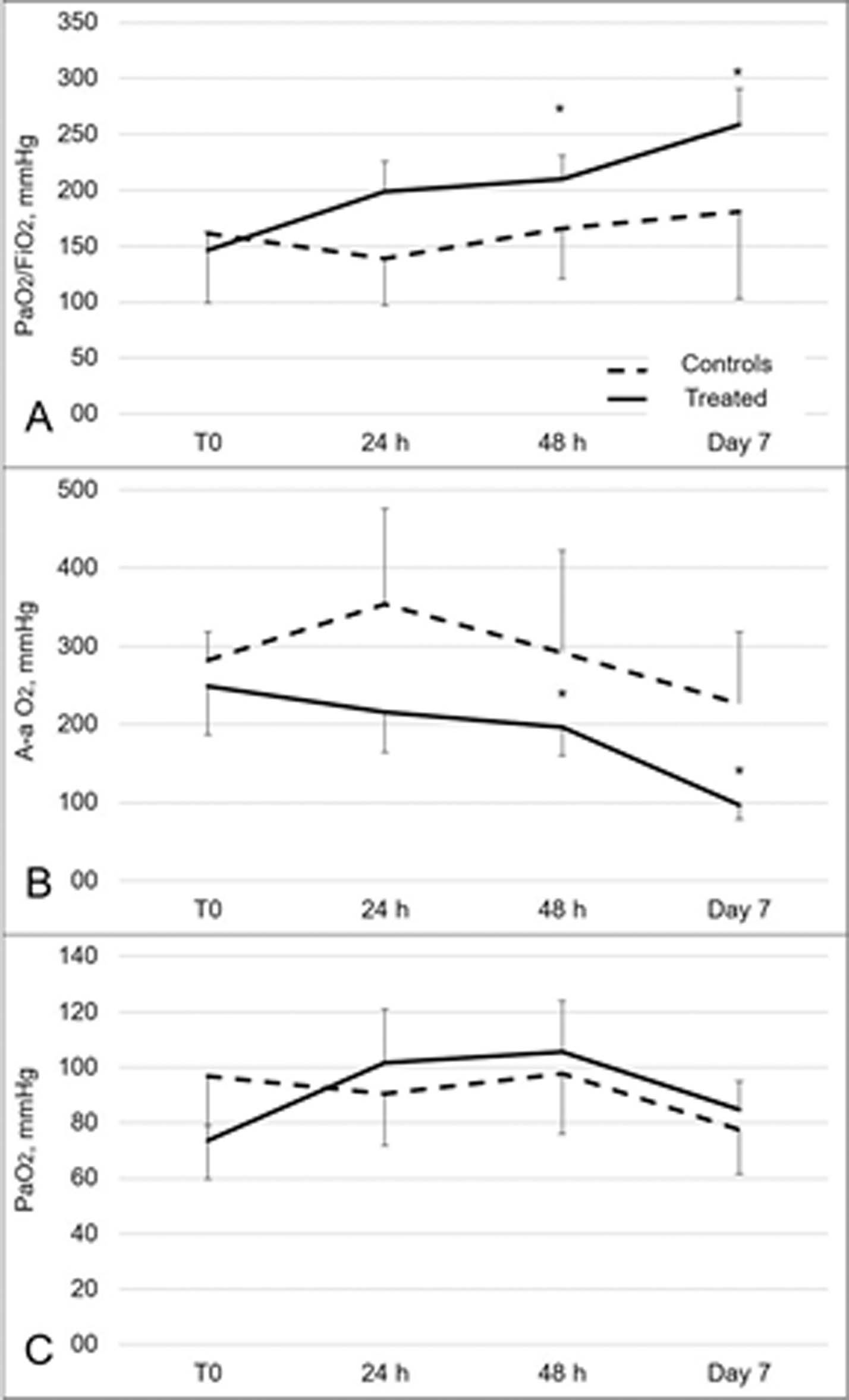
Fig. 1. Changes in oxygenation parameters in treated patients and controls. Changes in PaO2/FiO2 (Panel A), A-a O2 gradient (Panel B) and PaO2 (Panel C) after anti-platelet therapy administration are shown at different time-points. Values are reported as means and vertical bars are standard deviations. * pvalue < 0.05.
the first time that concomitant treatment with tirofiban, acetylsalicylic acid, clopidogrel and fondaparinux at a prophylactic dose could improve gas exchange efficiency increasing arterial oxygenation and A-a O2 difference in severe patients affected by Covid-19 with a thrombophilic profile. The patients enrolled in the present study had a median D-dimer of 3.375 μg/L, and a median SOFA score of 4, posing them at elevated risk of in-hospital death [17]. Despite patients’ baseline severity, the improvement in peripheral oxygen was consistently paralleled by a reduction in the respiratory assistance without major adverse events, supporting the idea that this treatment might be tested also in ICU patients.
Thrombophilia has been repeatedly reported in ICU patients with Covid-19 [3,4,18], often associated with dramatic increases in D-dimer and fibrinogen values [3]. Recent evidence further demonstrated that both D-dimer values >1.000 μg/L and higher SOFA scores were associated with increased odds of death in hospitalized patients with Covid19 [17]. Although sometimes DIC features have been observed in patients with severe Covid-19 [11], low platelets counts and bleeding were rarely reported [3,7,19], suggesting that hypercoagulability is fostered by the severe inflammatory state [3]. Electron microscopy and immunohistochemistry analyses have recently provided evidence of an elevated number of megakaryocytes and platelet-fibrin thrombi in alveolar capillaries from lung tissue of deceased patients with severe Covid-19 [6]. Megakaryocytes are hematopoietic cells specialized in the production of platelets that migrate from the bone marrow to the lung microvasculature [20], where they release platelets that are retained and sheared in the pulmonary capillary vessels [20,21]. We hypothesize that the administration of the GP IIb/IIIa inhibitor tirofiban might have prevented the formation of newly assembling clots promoted by the dysfunctional endothelium of lung capillaries. In fact, both hypoxia and sepsis can independently promote platelet aggregation [22], mediated by the release of Von Willebrand factor, which was found greatly increased in the whole blood of ICU patients with Covid-19 [3]. The latter effect, associated with the inhibition of cyclo-oxygenase (COX) present in platelets and megakaryocytes and promoted by acetylsalicylic acid and the clopidogrel-related modulation of the ADP megakaryocytes receptor [23], might have impeded the consolidation of forming thrombi, thus improving regional ventilation/perfusion mismatch and explaining the rapid increase in PaO2 and the reduction in A-a O2 gradient [24]. Moreover, it may be conceivable to suppose that the consequent reduction in the alveolar-endothelial thickness might have led to a local improvement in oxygen diffusion capacity [25] thus in part justifying the increase in arterial PaO2.
Platelet derived thromboxane A2 (TxA2) has vasoactive as well as pro-thrombotic properties, promoting the activation and aggregation of nearby platelets [26]. Moreover, it has been suggested that TxA2 may have a role in mediating the vasoconstrictor response of soluble fibrin in septic conditions [27]. Nonsteroidal anti-inflammatory drugs decrease platelet adhesiveness by interfering with platelet prostaglandin synthesis, such as the TxA2 metabolite TxB2 [26], partially restoring vasoplegic pulmonary regions and thus promoting an improvement in lung perfusion.
The tendency to normalization of the A-a O2 gradient couldn’t be secondary to recruitment or to changes in intrapulmonary shunt, as PEEP values (on average 10 cmH2O) were never increased during the study or were diminished, and CPAP was removed after 72 hours (Table S1 and Fig. 2). Indeed, an improvement in A-a O2 gradient could be secondary to an improvement in lung perfusion, with consequent positive effects on lung mechanics and respiratory drive, sparing patients from the harmful effects of excessive lung distension and mechanical stress [2]. It should be highlighted that participants entered the study at different timings in respect to the hospital admission. Despite a clear effect size gradient of the study therapy was not discernable, due to the natural history of the disease, it seems plausible to speculate that the maximal therapeutical advantage might be obtained in earlier disease stages. Recent evidence have linked myocardial injury and cardiovascular comorbidities with increased risk of severe disease and death [28]. In light of these data, we can not exclude that a precocious antiplatelet therapy might have a role in preventing cardiovascular complications in patients with COVID-19 pneumonia.
Currently, only one retrospective study demonstrated an advantage of anticoagulant therapy with low molecular weight heparin (LMWH) in terms of survival in patients with severe Covid-19 and signs of sepsisinduced coagulopathy [8]. Fibrinolytic agents such as tissue-type plasminogen activator have been proposed as a treatment for Covid-19 patients with coagulopathy [9]. However, we reason that an uncontrolled fibrinolysis affecting regionally inhomogeneous intra-capillary clotting might worsen blood arterialization by creating intra-parenchymal shunt when perfusion is restored in otherwise unventilated regions.
The present study has limitations. The results are influenced by a selection bias and by the limited sample size. Baseline characteristics and disease natural history varied from patient to patient, limiting the generalizability of the results. Considered the severity of the patients involved in the study, the case-control matching resulted difficult to perform. Thus the comparison might be biased by worse outcomes in the control group, which might be confounded by baseline lymphopenia, although the difference between groups was not statistically significant. However, it should be recognized that patients had a comparable mortality risk based on the severity of respiratory failure and the elevation of D-dimer. We are also aware of the multiple
Funzioni
La Fondazione
Legal Note